Chapter ten
Eye Evolution
Animation courtesy of
Daniel Cordier, CNRS, L’Université de Reims Champagne-Ardenne
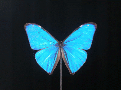
Iridescence: The observed color changes as a function of viewing angle. The colors of this Morpho butterfly range in wavelength from blue through purple to russet and walnut. The physical principle underlying this relationship between viewing angle and colors can be used to create an angular position detector. Such a device “reads-out” the angle to a target object as a function of the color impinging on a photosensor. One can construct such a detector using a thin film étalon. This essay explores the possibility that primitive directional eyes might have exploited this simple principle. The resemblance of the Morpho animation to a radar antenna is coincidental but in some sense, quite apt as a metaphor.
A walk in the park
Suppose one fine day on a walk in the park you find yourself paced by and then suddenly engaged in an argument with a madman. A day later, in reviewing the experience, you may discover that your own sensible, well reasoned arguments were somehow shaped and made strange by the surreal arguments of that madman. This is because either side of any argument is, in effect, a template that lends its shape to the opposite side of the argument.
If we practical, sensible everyday partisans of biology formulate arguments against the creationist template, we are basically arguing with a madman in the park. We should not be surprised to find that our own reasoning becomes, in this encounter, oddly distorted and not a little strange. Case in point. The wars of perfection, as follows:
The Wars of Perfection
Although the curious frontal wiring and backward looking orientation of the vertebrate retina is univerally accepted — the “why” of it has long since become a football in arguments between creationists and biologists. The argument seems to be framed around the question of perfection.
Biologists have observed that if the vertebrate eye were in fact intelligently designed, the vertebrate retina would be wired from behind. That way we would not have to peer at the world through the screen of the retina’s wiring system. There would be no blind spot where the optic nerve makes its exit passage through the retina.
It is urged that what we have here instead is in fact an example of “stupid design,” that is, a design which is not a very good one — imperfect in fact — and therefore cannot be defended as evidence of intelligent (perfect) handiwork.
The Imperfect Eye argument was conceived as an elbow jab at the creationists. Evolution, which is understood to be a rather accidental process, can produce imperfect results. This is okay, expectable, how it works. But to the creationists, who do not accept evolution, the imperfection of the vertebrate eye raises a problem, because in the biblical version of events, there were no mistakes.
Naturally, technically inclined creationists have countered with an earnest effort to show that the inversion of the retina is both intelligent and perfected after all.
This conflict over perfection, like any other argument about the existence or specific intervention of God, is just a pastime –a very human pastime. It is also a complete waste of breath.

If we are going to insist on imperfection, just to keep up our side of the argument against the creationists, we will end by obscuring an important fact: The level of structural precision evolution can produce is simply stunning.
The vivid blue of this Morpho butterfly is not pigment. It is a precision mirror that, at certain viewing angles, beams blue light into our eyes. The mirror is formed as a carpet of scales on the wings of the butterfly. Each scale is a multilayer mirror. Its multilayer structure is so fine that it is below the resolution limit of a light microscope. It can be made evident only with the help of an SEM.
In general, biological mirrors are formed from a stack of typically twenty reflective layers dimensioned and spaced – precisely — at intervals of a quarter-wavelength of light or at an odd multiple of a quarter-wavelength of light.
In multi-layer mirrors the evolutionary pressure is supplied by the physical principles of thin film interference.

This is an effect that comes into play when light waves are reflected from interfaces spaced apart by tiny, tiny distances — less than a few wavelengths of light. If you have ever spilled oil on water, or blown bubbles, you’ve seen this familiar effect.
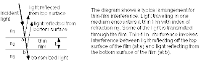
High light reflectance is a result of constructive interference. Light reflected from the top surface must interfere constructively with light reflected from the bottom surface, so the two waves must be in phase. The effect depends on the angle and wavelength of the impinging light, and on the exact dimensions and refractive indices of the structures the light penetrates.
In the butterfly’s wing scales constructive interference occurs only for odd multiples of a quarter wavelength (1,3,5) of light. Low reflectance, a result of destructive interference, occurs for even multiples. The principle of thin film interference is discussed in detail at the BU site.
The diagram above shows just two layers separated by a thin film. This structure can reflect only a few percent of the incoming light. Reflection owing to constructive interference can be enhanced to nearly 100% by stacking the mirror in multiple layers. In nature, we are talking about a biological structure with about twenty layers and gaps, dimensioned and positioned, accurately and repeatedly, at intervals on the order of an eighth of a micron.
Precision multilayer mirrors and gratings occur in the scales of butterflies and moths. Multilayer mirrors can also be found in the scales of other insects and fish, and in the tapetum lucidum of the eye. Insect scales in particular are hugely important in the history of science: Ernst Abbe’s theory of image formation is based on his insight, in 1872, into the diffraction effects of the multiple layers (multiple slits, viewed end-on) on light entering the imaging system of a light microscope through a butterfly’s scale. We owe our understanding of how images form — in cameras, in microscopes, in our own eyes — to an insect’s scale.
Here is some discussion of butterfly optics, structural color and natural photonics, on the site of the University of Exeter’s fascinating and impressive program on Natural Photonics. The best treatment I have seen of physics of the Morpho butterfly is on the site of Daniel Cordier.
Natural multilayer mirrors are typically reflective of colors, since they are dimensionally tuned to specific wavelengths. Note too that they will reflect different colors at different viewing angles. They are iridescent.
White light mirrors in nature, like fish scales (in the Herring, for example), are even more sophisticated. In the fish scale structure, three multilayer mirrors are overlaid. Each has a different natural wavelength. A third of the mirror reflects blue-green, a third reflects red purple, and a third reflects orange yellow. The net reflection of the stack is white light.
In sum these biological optical structures are almost crystalline in their, well — perfection. Examples of amazing structural precision – and movement — also abound at the level of molecular biochemistry and include nanomachinery like hemoglobin (the “molecule that breathes”) and the active site mechanisms underlying enzymatic reactions.
We can freely delight in our amazement, our scientific astonishment. Amazement does not compel us to fall on our knees. Nor is it embarrassing to be impressed by biological precision – as though we would somehow, by being impressed, give away points to the creationists on this absurd question of perfection.
The conflict between reason and faith antedates Darwin by about 15 centuries, and it cannot be “won” with Darwinian reasoning or any sort of reasoning at all. It is not a debate. It is a power struggle. Reason requires questioning — open, frequent, often defiant questioning. Faith is unquestioning acceptance and obedience. There is a good analysis of the origin and politics of this peculiar issue, which is a real quirk of Western civilization, in a 2004 book by the historian, Charles Freeman.
A short history of the vertebrate eye
In any event and in the meantime, the real question, which is not about perfection, has been pretty much left behind the door. Here is the question:
What is the evolutionary reasoning or narrative that accounts for the inverted structure of the vertebrate retina?
The question is typically answered in texts on the basis of ontogeny, and this is a safe and solid answer. But as usual there are riskier, more imaginative approaches with more style and promise. One proposal I have admired, because it is based on function, is that the modern vertebrate eye was once and in some sense still is a mirror eye.
 This type of eye uses a backwall mirror rather than a lens, or in combination with a lens, to form an image. The scallop eye is one modern example of a mirror eye. We may or may not have an interesting ancestor in common with the scallops, but in this hypothesis our eyes are at least analogous.
And the primitive chordate eye had no lens at all. The photoreceptors were positioned, aimed and wired to capture an image formed by the eye’s backwall mirror. This explains why the retina “looks backward” toward the mirror.
In a subsequent step, a lens evolved, perhaps to correct the optics of the backwall mirror. (This is thought to be the purpose of the lens in the eye of the modern scallop. It is shaped to eliminate the mirror’s spherical aberration.) Eventually the vertebrate lens took over the whole job of imaging, and the role of the backwall mirror shifted to other tasks. According to this hypothesis, the mirror on the backwall of the eye survives today in structures like the tapetum lucidum — the eye mirror that accounts for the shining eyes of cats by night. The cat’s eye shine helps its night vision, but a modern backwall mirror may be useful in other ways and in other creatures, including primates.
One can, of course, rewrite this narrative in many different ways. The one crucial element is the primitive chordate eye that worked — formed images — without a lens. The idea of a mirror eye as an ancestral eye is appealing because the thing evolves a retina and a lens in two distinct, successive steps. It is thus a plausible solution to the problem of simultaneity.
If we leave out the primitive mirror as a step in the evolution of the vertebrate camera eye it might seem necessary to evolve two very sophisticated and optically precise structures, the lens and the retina, one suspended above the other, each functionally dependent upon the other — simultaneously. The difficulty of imagining, let alone accomplishing a simultaneous evolution of the retina and the lens was first remarked by Darwin. The shortcut solution offered to us by the scallop is that it never happened. The retina appeared first. The lens evolved much later.
The computer model
There is a well accepted model for eye evolution which depicts the beeline evolution of a camera eye. (There is no evolutionary detour through a mirror eye or compound eye to arrive, much later, at a camera eye.) Although Darwin was stumped by the problem of simultaneity, it has proved possible after all to model with a computer a stepwise, linear process of evolving a camera eye with a lens positioned over a retina.
The starting point is a flat, trilayer sandwich comprising an outer protective layer (which is transparent), a layer of photoreceptive cells, and an underlying layer of pigment cells.
This is a minimum eye — a photosensor which, owing to the pigment backing, has directional sensitivity. Light can only reach it from one direction. When a shadow appears in that direction, the animal knows something’s coming, and from which way, and it can react directionally. Or, underwater, a directional eye can simply indicate which way is up by detecting the light of the sun.
From this starting point, the computer generates random deformations and selects for improving visual acuity. It turns out the structure evolves by first dishing to form a pigment eye cup. Then, ultimately, gradually, a lens is formed over the aperture.

While the eye is still an open cup, it functions as a directional sensor. Light impinging on a particular point inside the cup can only have come from certain directions. (A cup is more sensitive to direction than a simple flat sensor blocked, in one direction only, by a pigment layer.) To take full advantage of the directional quality of the cup, the interior becomes populated with photoreceptors. As with the mirror eye, one could remark that a retina emerges before the lens.
However, cupping does not really produce a nice imaging eye until the cup has become a sphere with a hole in it and that hole — now understood as an aperture — starts to close.
Two alternative paths to a camera eye
This is the second half of the eye’s story — after it has cupped beyond the hemisphere and begun to close to form an eyeball. The word camera means “room”, and a room implies enclosure.
In the limit of cupping, you would get a sphere with a pinhole aperture that produces a sharp but rather dim image on the retina, as in the eye of the modern nautilus.
One could imagine that in life, there would be at least two different possible sequences of evolutionary events as the aperture closes.
Perhaps the eye might close all the way to a pinhole aperture — and in this way discover the miracle of an image. And then invent the lens as a way to open the aperture up again — i.e, to capture more light without losing focus.
Or it could be the cupping process would not run to completion — that the closing aperture would be capped by a lens. An evolving lens would arrest the cupping process and produce an image before the aperture ever contracted to the diameter of a pinhole.
In any event: According to the model, using what we might call brute force evolution, it takes only about 400,000 generations, or perhaps a half-million years, to arrive at what we would regard as a modern camera eye. A half-million years is just a moment in geological time. In fact, over the eons, camera eyes have turned up many different times in creatures as diverse as spiders, squids and ourselves. From the short time frame it is easy to conclude that the camera eye has evolved several different times in several different species, each time starting from scratch. But this has turned out to be an incomplete or simplistic idea. The more we learn about development, and how to think about development, the more intricate this story seems to become.
The original computer model is a classical work (Nilsson D-E, Pelger S (1994) A pessimistic estimate of the time required for an eye to evolve. Proc R Soc Lond B 256: 53-58). I think what the model describes best is the most direct way to achieve a camera eye.
 Colorized SEM of a jumping spider, courtesy of Tina Weatherby Carvalho, Biological Electron Microscope Facility, U. Hawaii
The spider’s camera eye started out in a different direction — it was probably a compound eye once, and then its multiple, faceted lenses coalesced. Spiders have lots of eyes, primary and secondary, and their secondary eyes sometimes exhibit lens/mirror combinations. So the mathematical model does not really attempt to tell the spider’s complicated story. Nor was the model designed to be specific to vertebrate evolution. It does not offer any special explanation for the backward-looking retina of the vertebrate eye.
And we are left with some questions about the pull and tug between the evolving camera eye’s two distinctly different functions. Initially the modeled eye is a directional sensor. Ultimately it is an imaging eye. The transitional steps or logical bridges between these two different types of eyes — two different physiologies, really — are not clear to me. This is not a criticism of this model. It is a criticism of the way we think about the evolution of vision.
The imaging eye is the superstar. The directional eye is marginalized — we tend to fast forward past it on our way to the heroic, climactic formation of an image. This is perhaps an error in emphasis. Many of the parts and pieces of the imaging eye must have evolved in the service of a machine which could not and never did form an image, i.e., the directional eye. Among the parts and pieces I have in mind are the color photoreceptors and the backwall mirror.
Good enough now.
The hypothetical vertebrate mirror eye would have given the ancestral vertebrates a huge jumpstart on vision — a good, working imaging system mounted early in the game.
They probably needed a jumpstart. The invertebrate (arthropod) compound eye — was already advanced, perhaps perfected, in the lower Cambrian. There were chordates in the Cambrian, but they are thought to have been sightless. The vertebrate camera eye was a come-from-behind effort.
The earliest vertebrate camera eye for which there is fossil evidence appears to have been installed and working in a fish that lived about 430 millon years ago. By that late date the arthropods, with their compound imaging eyes, had already been looking around (for things to eat, such as the small, supposedly blind chordates) for about 100 million years.
A mirror eye is optically inferior to a modern eye with a fine converging lens. But it works and, more to the point, in evolutionary terms it works almost immediately. As soon as the primitive eye is cupped, the structural basis for a mirror is in place. 
Parabolic reflecting antennae, Imperial War Museum.
In 1940-41, in the urgency of the Battle of Britain, English radar scientists had a peculiar motto, intended to help restrain themselves from perfecting their instrument: “Good enough now.” They coined this little slogan because it was far more important to make radar work and put it into service immediately, even as a makeshift, than it was to make it perfect. “Good enough now” limns an evolutionary principle for the mirror eye.
When the lens finally evolved, optical excellence became possible. The mirror eye hypothesis clearly explains the backward-looking vertebrate retina: In the old days, there was something to see back there: The world was reflected in and focused by the backwall mirror. For our remote ancestor, the mirror eye would have been a timely solution and great thing. But it necessarily left the vertebrates a legacy of — and a problem with — light scattering by the retina’s own intervening wiring system.
Why a mirror? The color radar hypothesis.
To get a better sense of the prehistory of the vertebrate eye, we should reconsider the directional eye. 
This early eye is informally conceived by most of us as a “real” eyeball that is still under construction. (Informally, that is, because we relax the usual teleological objections.) The primitive eye might be flat, it might be dished, it might be half an eye, a hemispherical cup – but it could not yet form an image. So in what sense was the directional eye a finished product? What could it actually do?
The possibility exists that the directional eye was once a very finished product: a passive radar system based on the detection of color. Its structural basis would be a monolayer — and then later a multilayer biological mirror. With this type of directional eye, color is not perceived by the animal as painted onto a literal image, as it is with a modern camera eye. Instead color (wavelength) is read by the animal as a signal that reveals the solid angle to another, targeted animal – prey or predator. When the target animal moves, the detected color changes, and it changes in such a way as to suggest, to the beholder, the speed and direction of the targeted animal’s path.
We cannot know if a color-based radar eye existed 500 million years ago – or ever existed. It will always be a pure speculation. Probably best to just regard it as an eye that never was – an imaginative exercise. But if it did exist in the world, perhaps by the early Cambrian, it could have had a fairly long run.
 Photo of a Wiwaxia model courtesy of the Royal Tyrrell Museum in Alberta. The iridescence of the blades decorating this hatlike Cambrian creature is owing to a natural diffraction grating. The iridescent effect and its underlying physical structure were discovered by the Oxford zoologist, Andrew Parker, who recounted the discovery in his book, “In the Blink of an Eye.” Note that the blades, in effect, shoot colors in every direction. Why did this little Cambrian animal find it useful to bombard its immediate surroundings with iridescent colors? One possibility is that Wiwaxia’s blades evolved as a very effective radar jamming strategy.
In the end a color based radar eye could have been defeated by color itself — by radar jamming. Iridescent displays, which were popular in the Cambrian and later, could have flummoxed a color radar eye. We know of no modern examples of a radar eye, so if it ever existed, it was in fact defeated.
It could be an interesting high school science project to test the possibilities of this hypothetical but intriguing form of vision by actually fabricating a color-based radar eye using analogous optical, electronic and logic hardware. Better, it could be readily modeled or simulated with a computer program, perhaps Mathematica (TM) or a modified drawing program. In a virtual world one could try out the device and perhaps gradually re-invent it (as evolution would) to suit several different underwater settings, since wavelength and light intensity and directionality all change as a function of depth.
The solid angle problem
To see the basic problem facing a primitive directional vertebrate eye a little more clearly, let’s consider briefly a different, perhaps rival solution — a type of anatomical structure favored by bugs and other arthropods. Here is a compound eye. Notice that the lens/receptor units are arranged and packed into a convex hemispherical array. Each photoreceptor is aligned, rather like a telescope aimed at the sky, along a specific and unique solid viewing angle. In the most primitive, no-neck animals, the eye could not be quickly aimed at a target. The head did not move relative to the body. The eye did not move within a socket. The retina did not move within the eye. It was a question of aiming the whole animal. Not a problem for a modern, expertly piloted high speed insect like a housefly, for example — but not so easy for a soft, slow, primitive underwater creature, crouching on the seabed.
For a primitive fixed eye, one good solution is to collect information from an array of receptors arranged as a convex hemisphere, parsing the world into a global array of solid angles. Suppose one of the many photoreceptors reports a target – reflected light or shadow. The identity of that receptor, because of the direction in which it is structurally fixed and aimed, automatically defines and reports the solid angle to the target.
The arthropod’s convex hemispherical eye structure shows us a literal minded engineering solution to the problem of parsing the world into an array of solid viewing angles. The hypothetical color-based radar eye does the same job: It determines solid angles to a target – a point of light or darkness. It uses much less equipment, and it need not be hemispherical. It can be perfectly flat. But it has the same technical objective, which is to detect a change in the light at some specific solid angle drawn from the center of the eye.
How does a radar eye work?
Suppose we start with the familiar concept of “the minimum eye,” the primitive structure posited in 1994 by Dan-Erik Nilsson and Suzanne Pelger as a reasonable launch point for mathematically modeling camera eye evolution.
The minimum eye is a flat, trilayer sandwich comprising an outer protective layer (which is transparent), a layer of photoreceptive cells, and an underlying layer of pigment cells. This is a minimum eye just by definition – a photo sensor which, owing to the pigment backing, has directional sensitivity. Light enters from the front, but not from the back, where it is blocked by pigment. A binary device. Yes, no. Up, down. Nothing to it.
 But there is a wonderful built-in complication. As noted, the minimum eye is fitted with a transparent outer layer and across this layer, which is essentially an étalon, we must expect to observe the effects of thin film interference. The important physical property of the thin film sandwich structure, from the standpoint of building a directional eye, is not color per se. It is iridescence, that is, perceived color change as a function of viewing angle or, more importantly for our purposes — angle of illumination. The position and therefore the viewpoint of the photoreceptors is fixed by the anatomical structure of the eye. What can vary is the angle of the incoming light reflected from (or interrupted by) an object.
In the discussion of biological mirrors here, we have repeatedly remarked on their typical property of iridescence. Iridescence is a natural phenomenon in which a surface exhibits different colors depending on the observer’s viewing angle. A good example is the surface of a Morpho’s wing. If the Morpho has alighted and is sitting perfectly still — and you edge sidewise to shift your point of view a bit, the color reflected into your eye by the wing will be seen to change from blue to a russet or walnut color.
Slightly different angles produce distinctly different colors, in this instance, russet vs blue. Iridescent color varies with viewing angle and with the angle of illumination. Photo courtesy of Daniel Cordier, ENSCR, France

The optics are reversible. If your point of view is held fixed, and the butterfly moves, the color you see will change. The color you see will depend upon the solid angle that one might draw between your eye and the reflective surface.
The Iridescent Eye

The hypothetical radar eye of the predator (if you could be transported back to the pre-Cambrian world with your own imaging eye in order to see it) would itself be iridescent. This is the eye of a modern bottom dwelling coffinfish, from the collection of the Yale Peabody Museum. The eyes have iridescent flecks. If an iridescent eye did once exist, perhaps vestiges of its structure could still be identified.
In the simplest, earliest circumstance, the predator’s target would not be iridescent. It would simply be an object, possibly a living object, illuminated by ambient daylight underwater. Underwater lighting is a complicated story. For our purposes this object, a prey animal for example, can be imagined as either reflecting or interrupting a ray of light impinging on the eye. I find it easier to visualize the ray interrupted by a black object.
Suddenly, the watchful predator notices, blue light has disappeared. Gone soft or gone out completely. This means: an object exists somewhere in a sharply defined cone of possible solid angles, let’s say 35 degrees. But 35 degrees right or left? Fore or aft? And how far away? One eye can distinguish the presence of an intruder, but it can only report its solid angular position as a point existing on a conical plane of possibilities.
So it would take at least two flat, simple radar eyes to construct, in effect, a triangulating radar system – each eye, on the alert, perceiving the instantaneous weakening and strengthening of different colors.
As the object moves across the predator’s “field of view,” the spectrum of each eye trills colors, like running a finger up a keyboard. The spectral sequence shows direction. The rate and rate-of-change of the trill show speed and acceleration. Stereoscopic logic triangulates the instantaneous position.
But note clearly that the animal never “sees” color as we do. For this creature, color does not exist. We can say “trills colors” but the animal does not mutter to itself a litany of “red-orange-yellow-blue-indigo-violet” as an object traverses its field of view. Its photosensors simply detect the intensification of light at certain wavelengths at certain points. Detected bands of intensified light correspond, in turn, to certain solid angular positions.
The AND logic that puts the radar eye’s color information together would not be a bad prototype for the AND logic that is thought to support trichromacy in a modern, imaging eye.
Parts and pieces
What would it take to build a radar eye, structurally? Not much. It can be flat. In a pre-Cambrian and lower Cambrian world of flat creatures, so recently descended of flatworms – floating doormats, some of them — flatness is a style motif. The detection system needs a zero, that is, it must be oriented vertically, pointed “up.”
 Modern coffinfish, photo by the Yale Peabody Museum.
Modern fish do this with amazing accuracy, and it does seem that “up”, or “toward the light” is a concept in the repertoire of the most primitive sorts of creatures.
The minimum eye requires a light sensitive cell capable of color discrimination. It requires a doubly reflective layer, an étalon. The power of the reflector and, thus, the range of the eye could be improved by adding layers, producing a multilayer mirror, but a simple monolayer would do for a starter eye.
The photoreceptive cells are mounted in such a way that they “look at” the thin film reflector. Photosensitive elements could be opposed to the surface. They could be positioned in front of the étalon, or behind it, or they could penetrate into or interleave with transparent multilayers.
How can a primitive photoreceptor detect color? Color sensitivity can be provided to a monochromatic photoreceptor physically, by varying or dialing or oscillating the critical distance between the interfaces of the thin film. Chemical tuning to particular center wavelengths, as in a modern color photoreceptor, could evolve in due time.
When we look at a modern cone or rod cell, we immediately observe the orientation and spacing of the multiple disks. It could be that this bag-of-cookies structure is a relic of a photoreceptive system that was once embedded in, and integral with, the stratified mirror of an ancient radar eye.
In the Stiles Crawford Effect of the second kind, the color of monochromatic light is perceived to change as a function of its angle of incidence on the retina. It might be that this effect had a place or purpose in the prehistoric directional eye.
The radar eye, as an element we might wish to introduce into the dialog of eye evolution — is a splendid parts bin for the modern, imaging eye that would have rather abruptly succeeded it. The radar eye supplies a retina populated with color photoreceptors, retinal circuitry, a species of stereoscopic logic and – my personal favorite – a backwall mirror.
The end of an era
Why an abrupt shift to the imaging eye? Imagine an ocean full of edible and abundant iridescent creatures, beaming bright colors in every direction, colors evolved to jam the radar eye. It becomes more and more difficult for animals equipped with the radar eye to do well. The radar eye makes mistakes. The predator lunges in the wrong direction. The prey flees straight into a predator’s mouth. The directional eye isn’t really workable anymore.
Then imagine inserting into this environment a new kind of predator equipped with an imaging eye that can actually perceive, rather than simply detect — colors. An imaging eye is vastly superior to a radar eye.
Maybe she has a mirror eye, maybe she has a faceted compound eye. (We are guessing we are in the pre-Cambrian or lower Cambrian, but we don’t really know where we are in geologic time.)
We do know she is hungry. She looks around. So many brightly colored snacks. So easy to see, so easy to track. A massacre ensues. She thrives on easily-won protein. Before long the iridescent colors fade. If there are any fish around, then fish scales will tend to overlay, evolving from iridescent color to white reflecting invisibility. Camouflage becomes fashionable. And in the end everyone who is left who has eyes — has imaging eyes.
Eight steps
Let’s pull together these several threads of speculative ideas and see if we can synthesize a single narrative line — a sequence of evolutionary steps leading to the modern vertebrate eye.
Here again is our friend the scallop: hardly a vertebrate yet not very far along, perhaps, from the split between invertebrates and protochordates. Suppose we regard it as an eye in mid-passage, a snapshot exhibit of the technical features that may have succeeded each other in the course of the evolution of our own modern eye.
It was first discerned by Michael F. Land, and reported in 1965, that the scallop eye was forming an image with its mirror, rather than with its lens. (Land, M.F. 1965. Image formation by a concave reflector in the eye of the scallop, Pecten maximus. J. Physiol. (London) 179: 138-53.)
Mirror eyes are also described from page 104 in Animal Eyes, which the definitive book on the whole subject, written by Michael Land and Dan-Erik Nilsson.
The scallop eye has been presented here as a good example of a mirror eye, but we have been developing a novel sense of where that backwall mirror, and the retinal photoreceptors nested against it, might have come from.
1) Specifically, we can now guess that the mirror first arose as a single transparent layer, an étalon, originally used as a component of a solid angle detector. Its purpose was to convert the angle or slant of an incoming light ray into a detectable color.
2) To multiply the sensitivity of the étalon, and add range to the detector, the single layer evolved into a multilayer, a strong reflector of light into the photoreceptors and – thus — a pretty good mirror.
3) In the service of color detection, color sensitive photoreceptors and the retina evolved, firmly mounted against the surface of the mirror.
4) To improve the directionality of this purely directional eye, the tissue of the eye cupped and the backwall mirror curved. This helped in triangulating targets between or among two or more eyes, essential for rangefinding. Note that cupping the backwall changes the interference effects one would expect of a flat eye. The curvature of the backwall alters the incoming ray’s path length between layers as measured at different points along the backwall. This is an effect to compensate for or try to to use productively in modeling the system, perhaps as a means to create and detect rings.
The curved mirror and retina were already in place, according to this narrative, long before the evolving eye ever captured its first image. The animal could not “see,” in the sense of imaging, but it had a rather precise sense of its own position relative to predator or prey. This is the type of positional sense that one could get from radar set: An accurate detector reporting the target’s direction, speed, acceleration, and range. And perhaps size.
Then what?
To defeat this type of eye, there followed an age of iridescence, possibly in the Cambrian. Surrounded by color, drowning in color — the radar begins to jam. The animal makes costly mistakes.
5) A happy accident occurs. The retina actually doubles. A duplication, one of evolution’s favorite stunts. As in modern animals like the turtle, a layered retina appeared. Imaging is now possible, very close.
6) All it takes is a bubbling, a physical separation of the two retinas, in the manner of the scallop. And suddenly, voila, we have an imaging mirror eye. One retina, the new retina bubbled up away from the mirror, will capture an image focused by the backwall mirror. Another retina, the original backwall retina, remains stuck in place, plastered against the backwall. The old retina is not well defined in the 1906 Lankester drawing, but the retinal split it is described by Schwab in some detail.
There ensues a transitional period when the eye has two visual senses to process: a true image, from the upper retina; and the original color radar readout, from the lower retina. Ultimately the outboard, imaging retina triumphs, takes over, and we can have a true mirror eye.
7) A lens evolves to correct the spherical aberration of the backwall mirror. Now that the mirror is focusing an image, the aberration suddenly matters.
8) The lens can do more. The lens ultimately takes over the job of focusing an image on a retina.
But which retina? This is a guess I cannot make, but the end product is evident. Ultimately the retina in the middle of the eye migrated back “home” to the eye’s backwall. The evolutionary pressure to do this would be to improve and perfect the optics of a camera eye. In the end, the optics of the original mirror eye no longer matter. The mirror finds other work, as the tapetum lucidum.
Conclusion
I should probably re-emphasize the obvious — that this is a speculation. There is no other way to get at the problem. Fossils, however abundant, will never show us how eyes evolved. Eyes are soft tissue structures, rarely preserved. Comparative anatomy, engineering imagination, molecular clues, computer modeling and simulation are our best tools, and our best product will always be a good guess.
A footnote about Step 5, the doubling of the primitive retina to create a tiered retina. This is not a novelty in nature. Tiered retinas can be discovered in modern animals, usually where there exists no mechanical means for accommodation. Notice in the drawing, however, that in the scallop there are not only two retinas – there are actually two anatomically distinct optic nerves. When the two retinas re-married, as the lens took over from the mirror, the retinal structure on the backwall would still, for a while at least, represent a bipartite retina.
The bipartite retina is a concept we have repeatedly advanced, in this essay, as one likely means for separately and simultaneously detecting both the Fourier plane and the imaging planes of the lens in the vertebrate eye. It has been suggested that the Fourier plane or “back focal plane” is also the plane of memory.